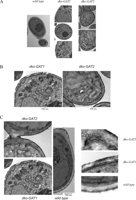
FIG. 9.
Identification of distinct membranous arrays and cortical ER alterations in dko-GAT1 and dko-GAT2 cells by electron microscopy.(A) Comparison of wild-type, dko-GAT1, and dko-GAT2 representative cells analyzed by electron microscopy. All cells were grown in rich medium containing galactose. (B) Enlargement of a portion of dko-GAT1 and dko-GAT2 cells showing aberrant membranous arrangements compared to those of the wild-type cells. (C) Detailed analysis of the cortical ER in all strains analyzed shows altered PM and cortical ER structural morphology in dko-GAT2 cells. Boxes indicate the areas that have been enlarged on the right, asterisks denote mitochondria wrapped by ER in dko-GAT1 cells, and arrows point to dilated ER in dko-GAT2 cells.

