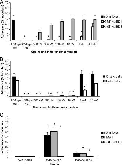
FIG. 4.
Inhibition of Hsf-mediated bacterial adherence by purified GST-HsfBD1 and GST-HsfBD2. (A) HeLa cell monolayers were preincubated with 0.1 to 500 nM purified GST-HsfBD1 or GST-HsfBD2 for 90 min. Subsequently, H. influenzae C54b−p−Hsf+ was inoculated onto monolayers, and adherence was measured with quantitative adherence assays. (B) Chang and HeLa cell monolayers were preincubated with 0.1 to 500 nM purified GST-HsfBD1 for 90 min. Subsequently, H. influenzae C54b−p−Hsf+ was inoculated onto monolayers, and adherence was measured with quantitative adherence assays. (C) HeLa cell monolayers were preincubated with 100 nM purified GST-HsfBD1 or purified HMW1 for 90 min. Subsequently, DH5α expressing the presentation vector alone, HsfBD1, or HsfBD2 was inoculated onto monolayers, and adherence was measured with quantitative adherence assays. In all panels, adherence is expressed as a percentage of the bacterial inoculum that bound to the epithelial cell monolayers. The bars and error bars indicate the means and standard errors of three measurements from representative experiments. In panel A, the asterisks indicate statistically significant differences (P < 0.05) for comparisons with H. influenzae C54b−p−Hsf+ with no inhibitor. In panel B, the asterisks indicate statistically significant differences (P < 0.05) for comparisons with H. influenzae C54b−p−Hsf+ with no inhibitor with Chang and HeLa cells. In panel C, the asterisks indicate statistically significant differences (P > 0.05) between the values indicated by the brackets (e.g., adherence by DH5α/HsfBD1 was statistically significantly different when monolayers were preincubated with no inhibitor versus GST-HsfBD1).

