
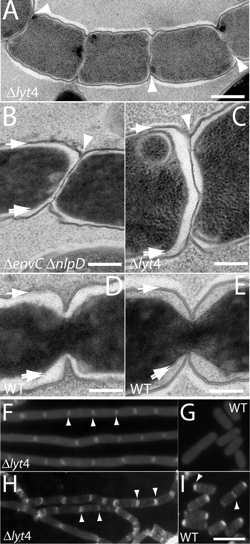
FIG. 5.
Septal PG splitting defect in mutants lacking LytM factors. TD22 (Δlyt4) (A and C), TB156 (ΔenvC ΔnlpD) (B), or TB28 (WT) (D and E) cells were mildly plasmolyzed and processed for EM (see the text for details). Shown are representative thin sections highlighting the septal PG splitting defect of TD22 and TB156 cells (A to C). The TB28 thin sections are provided for comparison to show that the constriction of the outer membrane and PG layer is normally tightly coordinated with the constriction of the inner membrane (D and E). The single arrows point to the outer membrane and PG layer surrounding the cell body. These two layers were not resolved at the cell periphery. The double arrows point to the inner membrane surrounding the darkly stained cytoplasm, and the arrowheads point to what appears to be an unsplit layer of septal PG in the chaining mutants. (F and G) Fluorescence micrographs of purified PG sacculi from TD22 (Δlyt4) (F) and TB28 (WT) (G) stained with amino-reactive Texas Red SE. (H and I) Fluorescence micrographs of purified PG sacculi from TD22 (Δlyt4) (H) and TB28 (WT) (I) stained with an Alexa 488-labeled lectin (WGA). Examples of intensely staining septa or poles are highlighted with arrow heads. Bars: 500 nm (A), 200 nm (B to E), 4 μm (F to I).