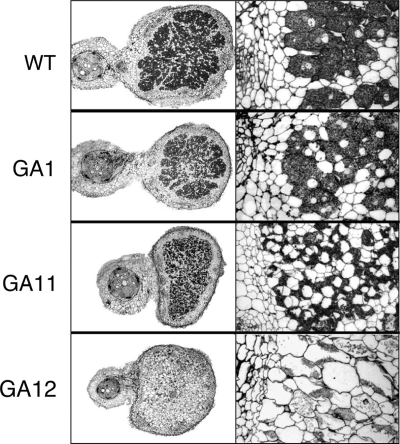
FIG. 4.
Histology of P. vulgaris nodules induced by R. tropici strains CIAT899 (wild type [WT]), GA1, GA11, and GA12 examined by light microscopy 20 days postinfection. Images shown on the left are cross-sections of whole nodules, and images on the right show more detail of internal regions of these nodule cross-sections at a higher magnification.

