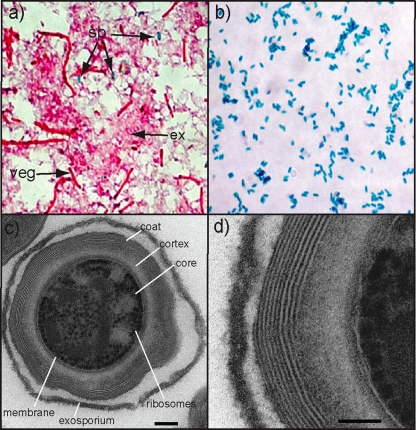
FIG. 1.
Visualization of pure C. difficile spores. (a) Endospore stain of C. difficile culture showing the pink vegetative cells (veg) and pink extracellular matrix (ex), with a few interspersed green spores (sp). (b) Purified C. difficile spores are stained green. (c) Transmission electron microscopy of sectioned C. difficile spores, demonstrating the spore ultrastructure including the exosporium, coat, cortex, core, membrane, and ribosomes. Bar, 100 nm. (d) Magnified section of outer surface of spore. Bar, 50 nm.

