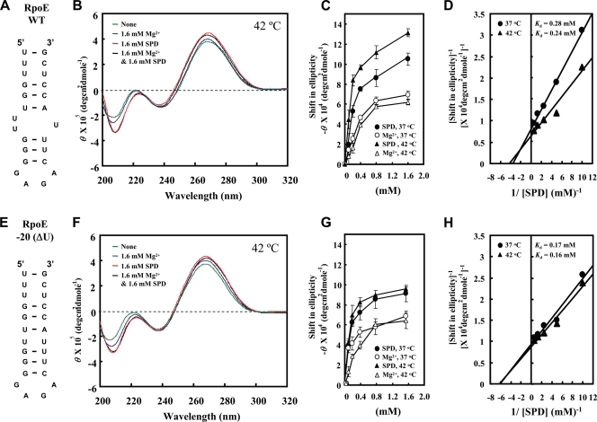
FIG. 6.
CD spectra of RpoE WT RNA and RpoE −20(ΔU) RNA. (A and E) Structure of RpoE WT RNA and RpoE −20(ΔU) RNA. (B and F) CD spectra were recorded as described in Materials and Methods. Green line, no addition; blue line, 1.6 mM Mg2+; red line, 1.6 mM spermidine; black line, 1.6 mM Mg2+ and 1.6 mM spermidine. (C and G) Concentration-dependent shifts induced by Mg2+ at 37°C, Mg2+ at 42°C, spermidine at 37°C, or spermidine at 42°C in magnitude at 208 nm are shown. Values are means ± standard deviations for three determinations. (D and H) The Kdapp values of spermidine for RpoE WT RNA and RpoE −20(ΔU) RNA at 37°C and 42°C were determined according to the double-reciprocal equation plot. SPD, spermidine.

