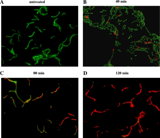
FIG. 5.
Fluorescence microscopy analysis of Syto 9/PI-stained strain SVMC28 Δsvl ΔlytA after MitC phage induction. SVMC28 Δsvl ΔlytA culture samples were collected at 40, 80, and 120 min after the addition of 0.1 μg/ml MitC, stained with a mixture of Syto 9 and PI, and visualized on a fluorescence microscope (magnification, ×630). As a control, the same cells were not treated with MitC. Different fluorescence patterns were clearly detected. (A) The untreated control corresponds mostly to bacteria exclusively stained with Syto 9. (B) After 40 min of phage induction, PI stained a few cells (dead cells), although the majority of cells stained only with Syto 9, indicative of intact membranes. (C) Eighty minutes after phage induction, almost half of the cells were stained with PI, with chains containing a mixture of PI- and Syto 9-stained cells. (D) After 120 min of phage induction, PI stained almost every cell. The chains showed few bacteria stained only with Syto 9. Each panel is from a representative experiment of four independent assays.

