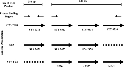
FIG. 1.
Schematic diagram of the genome organization of the loci used to design primers. The loci were from the three Salmonella strains S. Typhi CT18 (STY CT18), S. Paratyphi A (SPA), and S. Typhi Ty2 (STY Ty2). The expected size of the PCR product is shown at the top of the figure. Solid black arrows depict the position and direction of primer binding sites of the two sets of primers. The genome organizations of the three serovars are depicted as solid arrows, each representing a gene with its GenBank locus tag number given below. Broken lines represent genes that are absent in a particular serovar.

