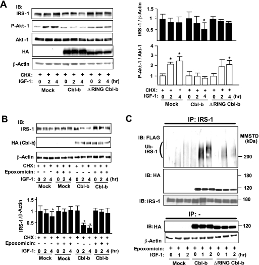
FIG. 3.
Transient expression of Cbl-b induces ubiquitination and degradation of IRS-1 in vitro. (A) COS7 cells were transfected with mock vector/pCEFL-Cbl-b-HA/pCEFL-ΔRING-Cbl-b-HA, pcDNA3-FLAG-Ubiquitin, and pLX2-IRS-1. To analyze the degradation rates of IGF-1 signaling molecules, COS7 cells were treated with 100 μg/ml cycloheximide (CHX) 3 h before treatment with recombinant human long-R3 IGF-I (10 ng/ml). Whole-cell lysates before or after IGF-1 treatment were subjected to immunoblotting (IB) for the indicated proteins. Densitometric ratios of IRS-1 to β-actin or P-Akt-1 to Akt-1 in the indicated samples are expressed relative to values at time zero of IGF-1 treatment. Data are means ± standard deviations (n = 3). *, P < 0.05 compared to the values at time zero. (B) Mock vector- or pCEFL-Cbl-b-HA-transfected COS7 cells were treated with vehicle (dimethylsulfoxide) or 100 nM epoxomicin in addition to CHX 3 h before IGF-1 treatment. Whole-cell lysates were subjected to IB for the indicated proteins. Data are the densitometric ratios of IRS-1 to β-actin in vehicle- and epoxomicin-treated cells. Data are means ± standard deviations (n = 3). *, P < 0.05 compared to the values at time zero. (C) Epoxomicin was added to all cells 3 h before IGF-1 treatment. Lysates from IRS-1-, FLAG-ubiquitin-, and Cbl-b-HA/ΔRING-Cbl-b-HA-expressing COS7 cells treated with IGF-1 for the indicated time intervals were immunoprecipitated (IP) with an anti-IRS-1 antibody. The immunoprecipitates were subjected to IB for the indicated proteins. Three independent experiments showed similar results. MMSTD, molecular mass standards.

