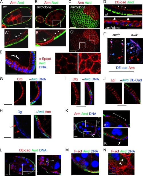
FIG. 3.
awd mutant clones show a depolarized accumulation of adherens junction components and epithelial abnormalities. (A) Stage 9 wild-type (wt) egg chamber doubly stained for Awd (green) and Armadillo (Arm; red). (A′) The apicolateral membrane localization of Arm is highlighted (*). (B) awd mutant clones (arrow in B′) in a stage 8 egg, revealed by a lack of Awd staining (green). These mutant cells show an overaccumulation of Arm (red; arrow), while neighboring awd+ cells show a typical apicolateral localization (*). (C) Surface optical cross-section of a stage 8 egg, showing double staining for Awd (green) and Arm (red). (C′) Staining of Arm alone. Arm overaccumulates on the awd mutant cell cortex. Close-up views show normal (left) and mutant (right) cells. Mutant cells contain an abundance of peripheral Arm and Arm-containing intracellular vesicles. (D) Enlarged views of a wild-type stage 8 egg chamber (top) with DE-cadherin (DE-cad) (red) at the apicolateral cell junctions (*). In the mutant clone (bottom), DE-cadherin spreads to the apical and lateral sides (arrows). Awd, green; To-Pro, blue. (E) Stage 10 egg chamber showing overaccumulation of α-spectrin (α-Spect) and a mild piling-up phenotype (brace). (F) Close-up views of awd+ and awd− stage 9 follicle cells, labeled for DE-cadherin (blue) and Arm (red). In the awd+ cell, besides colocalization at the apicolateral adherens junctions (*), separate DE-cadherin and Arm are observed on the lateral cortex (blue and red arrows, respectively). In the awd− cell, DE-cadherin and Arm colocalize to the apical membrane. In addition, cytoplasmic vesicles containing DE-cadherin (blue arrows), Arm (red arrows), or both (pink arrow) are identified. (G) A stage 7 egg chamber containing awd clones is stained for Crb, Awd, and DNA. Crb shows mild upregulation. (H) A stage 8 egg chamber containing awd clones is stained for Dg (blue), Awd (green), and Arm (red). Dg staining reveals a disrupted basal membrane (arrows), but the basal localization is not altered. (I) Stage 8 egg chamber stained for Dlg (red), Awd (green), and DNA (blue). (J) Stage 8 egg chamber stained for Lgl (red), Awd (green), and DE-cadherin (blue). Both basolateral markers show little change. (K) Stage 8 egg chamber showing stretched follicle cells (arrow in K′) and break-up of the epithelial sheet (bracket in K″). (L) Degenerating stage 9 egg chamber showing break-up of the epithelial sheet (bracket in inset) and severe piling up of follicle cells (arrow in inset). DE-cadherin, red; Awd, green; To-Pro, blue. (M and N) Stage 9 egg chambers containing awd clones are stained for F-actin (F-act) (phalloidin, red), Awd (green), and DNA (blue). (M) Adenoma-like epithelial expansion is observed. (N) In the most severe phenotype, large regions of the epithelium become multicell layered and invade deep into the germ cell complex (arrowheads). Bar, 20 μm.

