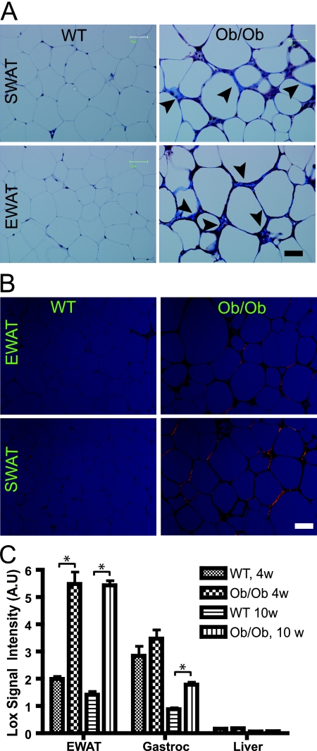
FIG. 4.
Fibrosis in dysfunctional white adipose tissue. (A) Masson's trichrome staining of SWAT and EWAT from 8-week-old wild-type mice (WT) and ob/ob FVB mice. Fibrillar collagens, primarily collagen I and III, are stained with blue, as indicated with arrowheads. Nuclei are stained with deep purple, whereas keratin stains red. Bar corresponds to 50 μm; three mice/group. (B) Picrosirius red staining of SWAT and EWAT from 8-week-old wild-type and ob/ob FVB mice. Picrosirius red was visualized under polarized light and shows collagen I in orange and collagen 3 in green. (C) LOX expression in the EWAT, gastrocnemius (Gastroc), and liver for wild-type and 10-week-old (10w) ob/ob C57/B6 mice, measured by the microarray analysis (five mice/group). Panel C was analyzed by Student's t test. *, P < 0.05; 4w, 4 week old; A.U, arbitrary units.

