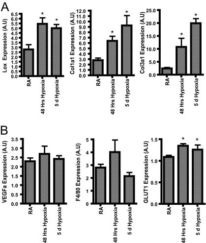
FIG. 8.
Respiratory hypoxia. Expression levels of LOX, Col1a1, Col3a1, GLUT1, F4/80, and VEGFa in the SWAT (A) and muscle (B) in mice breathing ambient air or 10% O2 for 48 h and 10% O2 for 5 days, as measured by quantitative RT-PCR. All data were analyzed by Student's t test, with four mice/group. *, P < 0.05; A.U, arbitrary units.

