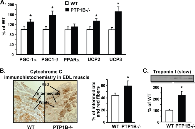
FIG. 2.
Effect of PTP1B deficiency on gene expression and fiber type composition in EDL muscle. (A) Levels of PGC-1α, PGC-1β, PPARα, UCP2, and UCP3 mRNAs were measured by quantitative RT-PCR. (B) Left, cytochrome c immunohistochemical staining in EDL muscle; right, quantification of red and intermediate fiber composition determined by cytochrome c staining in EDL muscle. (C) Immunoblotting of the slow-twitch type I fiber marker troponin I (slow) in muscle. All experiments were carried out using 3- to 4-month-old male mice on a chow diet. Data are expressed as means ± SEM; n = 6 to 8 for each genotype for panels A and C and 5 per genotype for panel B. *, P < 0.05 compared to results for WT mice.

