Figure 1.
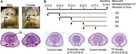
The time window for external genital masculinization. A, Sexual dimorphisms of the external genitalia in newborn mice (postnatal d 0). In males, the fusion of the urethral folds in the ventral midline results in canalization of the urethral epithelium (arrowheads) and bilateral fusion of the prepuce. The prospective corporal body (CB) condenses and bilaterally separates in the male but not in the female GTs. In the male, the anogenital distance, the distance from the external genitalia to the anus, is longer than that in the female (arrows). *, Urethral meatus. B, The timeline for flutamide treatment (upper panel). The GTs of all male embryos treated with flutamide on E15.5–16.5 and E16.5–17.5 (red lines) are demasculinized, displaying a morphology similar to those of the female GTs. The representative demasculinized and masculinized GTs and the GT of control mice with the vehicles are shown.

