Figure 6.
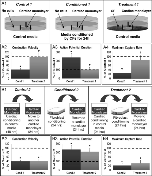
Effects of CF-conditioning in the presence of cardiomyocytes or cardiac conditioned media. (A1) Experimental setup. Control 1, a cardiac monolayer exposed to control medium for 24 h. Conditioned 1 (Cond 1), a cardiac monolayer exposed to CF-conditioned media for 24 h. Treatment 1, a cardiac monolayer surrounded by cardiac fibroblasts (without direct contact) and exposed to control media for 24 h. (A2–A4) Electrophysiological parameters in Cond 1 and Treatment 1, shown relative to Control 1 (dashed line) group. n = 12 monolayers per group. (B1) Experimental setup. Control 2, a cardiac monolayer exposed for 24 h to media conditioned by another cardiac monolayer for 48 h. Conditioned 2 (Cond 2), the same as Condition 1 in A1. Treatment 2, a cardiac monolayer exposed for 24 h to media conditioned for 24 h by CFs after being conditioned for 24 h by a cardiac monolayer. (B2–B4) Electrophysiological parameters in Cond 2 and Treatment 2, shown relative to Control 2 (dashed line) group. n = 12 monolayers per group. Asterisks denote significantly different results from control, while hash symbols denote significantly different from conditioned.

