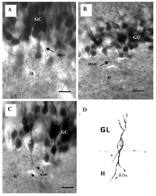
Fig. 2.

Two examples of biocytin-labeled granule cells with hilar basal dendrites from ischemic rats. A shows many labeled granule cells (GC) and one of these shows a large hilar basal dendrite (arrow, HBD) that enters the hilus (H). B and C show another example of a granule cell with a hilar basal dendrite (arrows, HBD). Note that this HBD is thick and it branches in the hilus (H). D is a camera lucida drawing of the same granule cell (*) with a hilar basal dendrite in C (asterisk). The basal dendrites (BDs) extend into the hilus while a thinner process arising from the base of this cell is the axon (a). Scale bars in A,C = 10 μm and B = 25 μm.
