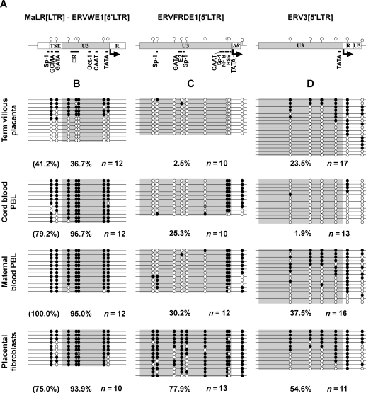
Figure 3.
CpG methylation envelope-coding HERV 5′LTRs in placenta-associated tissues. (A) Schematic representation of MaLR[LTR]–ERVWE1[5′LTR] (as in Fig. 2A), ERVFRDE1[5′LTR] and ERV3[5′LTR] analyzed regions. LTR regions are represented by boxes and CpG dinucleotides by circles on vertical bars. The U3 region (light gray) constitutes the retroviral promoter, transcription starts at the U3/R boundary (arrow). Putative transcription factor binding sites proximal to or overlapping CpGs are indicated. Downstream horizontal bars are provirus internal sequence. (B–E) CpG methylation of (B) MaLR[LTR]–ERVWE1[5′LTR] (as in Fig. 1), (C) ERVFRDE1[5′LTR] and (D) ERV3[5′LTR]. Methylation was determined by bisulfite sequencing PCR in villous trophoblast of term placenta, related fetal and maternal blood cells and in placental fibroblasts from chorionic villi of a first trimester placenta. Each sample result originates from the same conversion reaction. Each line represents an independent clone as determined by methylation and/or conversion differences. Methylated CpG are schematized by black circles, unmethylated CpGs by white circles and CpGs with undetermined methylation state by gray circles. Global methylation percentage values in the U3 regions (highlighted in gray) as well as in the MaLR[LTR] (in parentheses) are given below the respective area for each sample.

