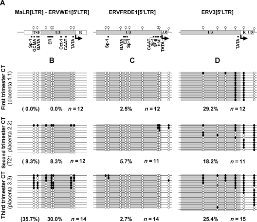
Figure 4.
CpG methylation dynamics of envelope-coding HERV 5′LTRs in cytotrophoblasts during pregnancy. (A) Schematic representation of the MaLR[LTR]–ERVWE1[5′LTR], ERVFRDE1[5′LTR] and ERV3[5′LTR] analyzed regions. (B–D) CpG methylation of (B) MaLR[LTR]–ERVWE1[5′LTR], (C) ERVFRDE1[5′LTR] and (D) ERV3[5′LTR]. Methylation was determined by bisulfite sequencing PCR in cytotrophoblasts (CT) at different times of gestation. One sample is represented here for each trimester i.e. CT of first trimester placenta from legally induced abortion (placenta 1.1), second trimester placenta from trisomy 21-affected pregnancy (T21, placenta 2.2) and term placenta from healthy mother (placenta 3.3). Each sample result originates from the same conversion reaction. Each line represents an independent molecule. Methylated CpGs are schematized by black circles and unmethylated CpGs by white circles.

