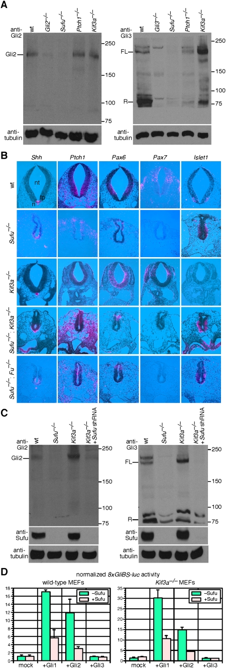
Figure 2.
Mouse Sufu regulates Gli protein levels independent of the primary cilium. (A) Western blots of lysates derived from wild-type (wt), Gli2−/−, Gli3−/−, Sufu−/−; Ptch1−/− and Kif3a−/− MEFs probed with anti-Gli2 and anti-Gli3 antibodies. Endogenous Gli2 and Gli3 protein levels (including full-length Gli3 and Gli3 repressor) are greatly reduced in the absence of Sufu. Both full-length Gli3 and Gli3 repressor can be detected in Ptch1−/− albeit the Gli3 repressor level is reduced. The ratio of full-length Gli3 to Gli3 repressor is altered in Kif3a−/− MEFs as reported previously (Liu et al. 2005). Gli2 processing is known to be extremely inefficient, and the Gli2 repressor form cannot be readily detected without additional enrichment steps using specific Gli-binding oligonucleotides (Pan et al. 2006). We also cannot accurately assess the full-length to repressor ratios for Gli2 and Gli3 in Sufu mutants. Tubulin was used as the loading control, and numbers on the right indicate locations of protein size standards. (FL) Full-length; (R) repressor. (B) Isotopic in situ hybridization using 33P-UTP-labeled ribo-probes (pink) on paraffin sections of wild-type (wt), Sufu−/−, Kif3a−/−, Sufu−/−; Kif3a−/−, and Sufu−/−; Fu−/− mouse embryos at 9.5 dpc. Loss of Sufu resulted in global up-regulation of Hh signaling and ventralization of the neural tube. Shh, whose expression is restricted to the notochord and floor plate in wild type, is extended dorsally in the absence of Sufu. Similarly, Hh target genes such as Patched 1 (Ptch1) are expanded dorsally, suggesting ventralization of the neural tube. The expression domains of neuronal progenitor markers (Class I genes, including Pax7 and Pax6, repressed by Hh signaling and Class II genes, including Nkx6.1, Nkx2.2, and Foxa2, activated by Hh signaling) are shifted. For instance, Pax7, the dorsal-most marker, is not expressed, and Pax6 expression is confined to the dorsal neural tube of Sufu−/− embryos. Dorsal expansion of Nkx6.1, Nkx2.2, and Foxa2 was also observed in the absence of Sufu (data not shown). Similarly, the expression domains of markers for differentiated interneurons and motoneurons are changed. For instance, Sufu−/− neural tube displayed dorsal expansion of Islet1 and Oligo2 (data not shown), which label motoneurons. By comparison, marker analysis revealed a partially dorsalized Kif3a−/− neural tube. The neural tube defects in Sufu−/−; Kif3a−/− or Sufu−/−; Fu−/− embryos resemble those in Sufu−/− mutants. (n) Notochord; (fp) floor plate; (nt) neural tube. (C) Western blots of lysates derived from wild-type, Sufu−/−, Kif3a−/− MEFs and Kif3a−/− MEFs expressing Sufu shRNA probed with anti-Gli2 and anti-Gli3 antibodies. Efficient Sufu knockdown in Kif3a−/− MEFs was verified by anti-Sufu antibodies. Gli2 and Gli3 protein levels are reduced in Kif3a−/− MEFs expressing Sufu shRNA to the same extent as in Sufu−/− MEFs. (D) Hh reporter assays using the 8xGliBS-luc reporter in wild-type and Kif3a−/− MEFs. Expression of Gli1 or Gli2 (but not Gli3) activates the Hh reporter, and Hh pathway activation is repressed when Sufu is coexpressed. Gli2 is known to activate Hh reporters less efficiently than Gli1 (Gerber et al. 2007). Loss of the primary cilium in Kif3a−/− MEFs does not impair Sufu's ability in repressing Gli-mediated Hh pathway activation. Error bars are standard deviation (s.d.).

