Figure 6.
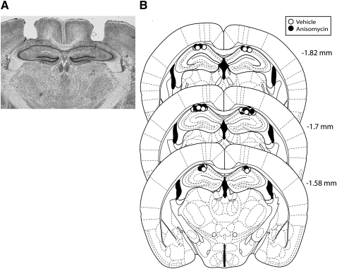
Hippocampal cannula placements. (A) Photomicrograph depicting a representative sample of an accurate bilateral dorsal hippocampal injector placement. The coronal slice was stained with cresyl violet and was taken from −1.7 mm bregma. (B) Actual injector tip placements in each animal used in experiment 5.

