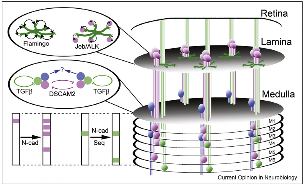
Figure 2.
The anatomy of ganglia, columns and layers in the Drosophila visual system. Photoreceptor axons (green) extend into the brain from the retina, with R1-R6 axons terminating in the lamina, and R7 and R8 axons terminating in the medulla. Within the lamina, R1-R6 axons choose targets arranged in a precise relative pattern, and make synapses with lamina neurons (magenta) arranged in an array of columns. These targeting choices are dependent upon afferent–afferent interactions mediated by the non-classical cadherin Flamingo, as well as by anterograde signals transmitted by Jellybelly (Jeb) and its cognate receptor, the tyrosine kinase ALK. Within the medulla, R7 and R8 axons make synaptic connections with medulla neurons (blue; only one type is shown for simplicity) arranged in columns. Projections from lamina neurons and photoreceptors are restricted to specific columns, and are laminated. Inhibitory interactions mediated by DSCAM2 serve to restrict a specific subset of lamina neurons to their target columns, while autocrine signals provided by TGFβ play an important role in tiling R7 terminals. As-yet unknown mechanisms presumably tile medulla neuron processes. The layer-specific connections of lamina neurons in the medulla are dependent upon the classical cadherin N-cadherin (N-cad), and the layer-specific connections of R7 and R8 axons are dependent upon a temporal control mechanism that utilizes a dynamic pattern of expression of the transcription factor Sequoia (Seq) to regulate the responsivity of growth cones to N-cadherin.

