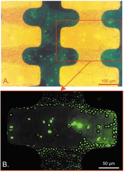
Fig. 2.

Views of a culture of strain T9/94 RC17 P. falciparum-infected erythrocytes containing approximately 1.1% parasitised cells suspended in 8.5% sucrose + 0.3% dextrose suspending medium on an interdigitated electrode with an applied field of 5 Vp-p at 200 kHz. Approximately 5 × 10⁶ erythrocytes were injected into the chamber. Both low-level bright field and epifluorescence illumination were provided. (A) Cells containing parasites exhibited a green fluorescence due to uptake of the potentiometric dye DiOC6 (3) into the parasite interior from the suspending medium and show brightly in the figure. (B) A magnified view under epifluorescence illumination confirmed that 95% of parasitised cells were repelled from the high field regions and could be washed free by flowing suspending medium.
