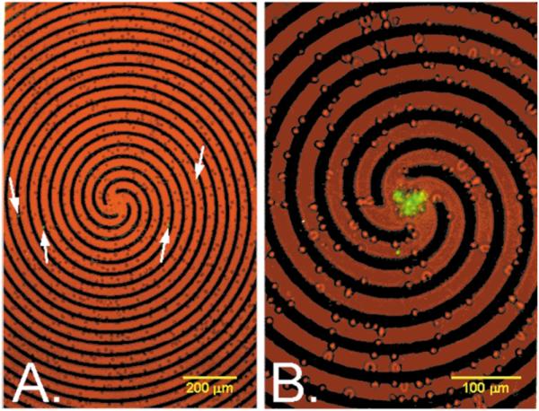
Fig. 3.

Erythrocytes containing approximately 5% parasitised cells on a spiral electrode array under the same suspending medium and staining conditions used in Fig. 2. Both low-level bright field and epifluorescence illumination were provided. (A) Prior to the application of a travelling electrical field, parasitised cells (arrows) were spread throughout the sample. (B) Application of four phase signals to the spiral electrode elements (3 Vp-p, 2 MHz) caused normal erythrocytes to be trapped at the electrode edges while parasitised cells were levitated and carried towards the centre of the spiral by the travelling field.
