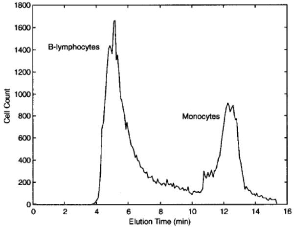
Figure 4.

DEP-FFF fractogram showing the separation of monocytes from B-lymphocytes. The injection and withdrawal syringe pumps were operated at 2 and 1.9 mL/min, respectively. Identification of monocytes and B-lymphocytes by flow cytometry was made possible by prelabeling them with PE-CD14 and FITC-CD19 antibodies, respectively. The cell suspension and DEP field conditions were the same as in Figure 3, except that the DEP field was swept between 20 and 40 kHz.
