
FIG. 3.
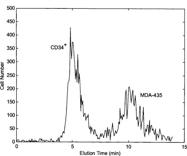
DEP-FFF fractograms for separating MDA-435 cells from CD34+ cells using the trap-and-release protocol. DEP field was operated at 40 kHz for 7 min and switched to 5 kHz for 7 min. CD34+ cells were prelabeled with PE-conjugated CD34 antibodies and were identified by flow cytometer to elute the chamber earlier than MDA-435 cells. DEP signal voltage and fluid-flow conditions were the same as those used for Figure 2.