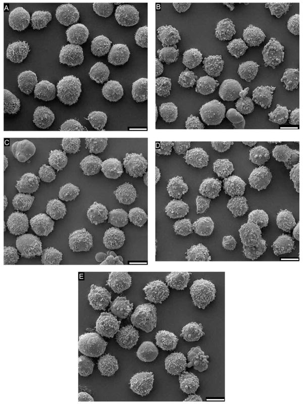
Fig. 6.
Scanning electron micrographs of HL-60 cells showing the appearance of untreated cells in comparison to treated cells at the times that toxicant exposure is first detected by the DEP responses. (A) Untreated cells at 30 min. (B) paraquat 19.44 μM at 15 min. (C) Styrene oxide 0.1 mM at 15 min. (D) NMU 0.2 mM at 30 min. (E) Puromycin 0.1 mM at 30 min. While most untreated cells exhibit complex surface morphology with relatively extensive microvilli, a few demonstrate a combination of coarse microvilli, blebs and smooth surfaces. In contrast, treated cells have generally fewer microvilli, more morphological irregularities, larger areas of smooth surface, significantly more blebbing and occasional apoptotic bodies. Bar length = 10 μm.

