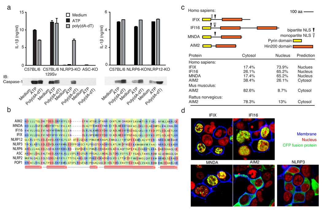
Figure 1. poly(dA-dT)-induced inflammasome activation.
a, LPS-primed macrophages from wild type or inflammasome deficient mice were stimulated as indicated and supernatants examined for IL-1β by ELISA or for cleaved caspase-1 by immunoblot. b, A multiple sequence alignment of human PYHIN and select NLR PYD domains, c, Domain structures of human PYHINs, with predicted nuclear localization signals and subcellular localizations (lower panel). d, Subcellular localization of CFP-tagged IFIX, IFI16, MNDA, AIM2 or NLRP3 (green) in 293T cells. Fluorescent choleratoxin stained membranes (blue) and DRAQ5-stained nuclei (red). Data from one experiment of three is shown (a, d).

