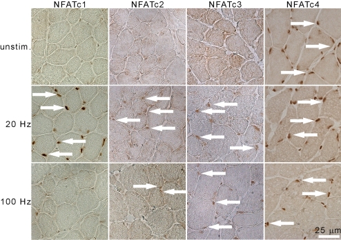
Fig. 5.
Nucleocytoplasmic shuttling of NFAT isoforms in response to electrical activity. IHC staining of endogenous NFAT isoforms in cross-sections of EDL electrically stimulated with patterns of impulses at 20 and 100 Hz as indicated, for 2 h. Contralateral EDL from the anesthetized animal was used as unstimulated control (unstim). White arrows indicate NFAT-positive nuclei.

