Fig. 3.
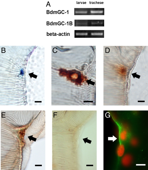
BdmGC-1 is expressed at main tracheal junctions. (A) Transcripts of BdmGC-1 and -1B expressed in third instar larvae and trachea. (B) RNA labeling with anti-sense RNA probes showing that BdmGC-1 transcripts are located at sites coincident with Inka cells at junctions along the main tracheal trunks. Specific Dig-labeled probes derived from the ECD region of BdmGC-1 were used for mRNA detection. Visualization is via alkaline phosphatase. (C) Inka cells exhibit strong ETH1-immunoreactivity, visualized with HRP-DAB. (D and E) BdmGC-1 signals visualized at junctions of tracheae using BdmGC-1 C-terminal-specific antibodies and BdmGC-1 N-terminal-specific antibodies, respectively, visualized by HRP/DAB. (F) N-terminal-specific antibodies neutralized by peptide antigen as control. (G) Merged view of fluorescent labeling of BdmGC-1 and propidium iodide counterstaining. (Scale bar, 20 μm.)

