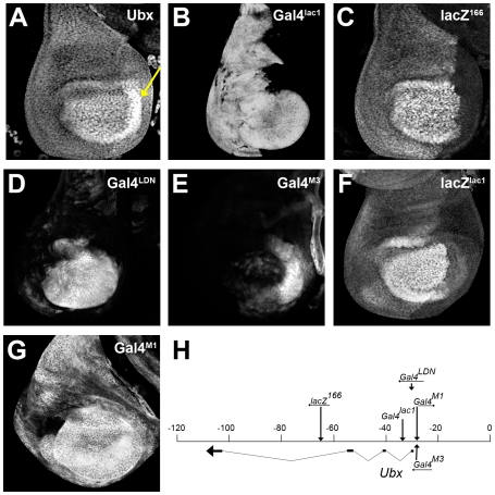
Figure 1. Ubx enhancer traps.
(A) Haltere disc stained for Ubx protein. Note the higher levels in the center of the disc and in the P compartment (arrow). (B–G) Patterns of Ubx enhancer trap expression in wild type haltere discs. The Gal4 inserts were monitored using a UAS-GFP transgene. (H) Map of the Ubx locus showing the location of the Ubx enhancer traps as described previously [28],[29],[30].

