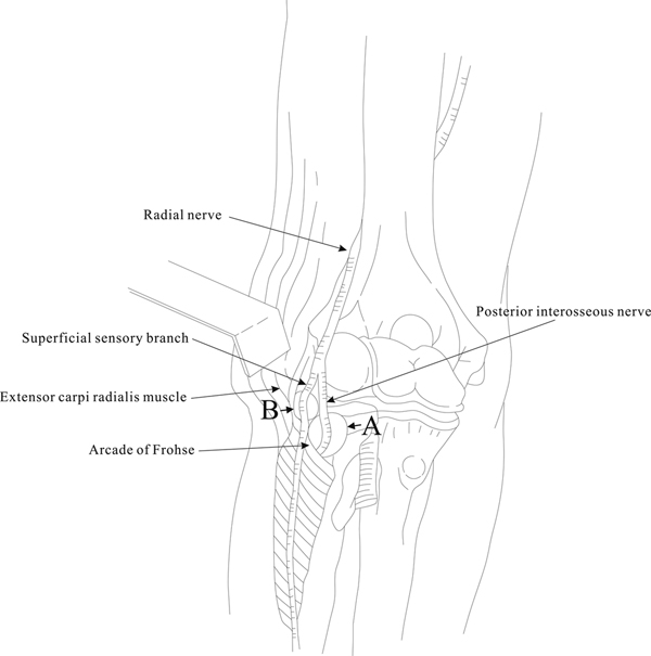
Figure 3.

Diagrammatic view of the radial tunnel showing compression of the ganglion on various locations of the branches of the radial nerve. Arrow A indicates the compression present in patient 1 and arrow B indicates the compression present in patient 2.
