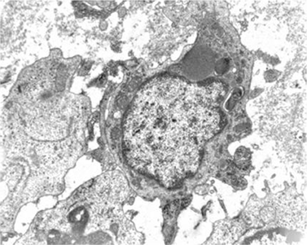
Figure 3.

Electronic microscopy of the excised tumor. Histiocytic lesion: giant cell with irregular cytoplasmic sprouts. Erythrophagocytes with large mitochondria, lysosomes, rough endoplasmatic reticulum and some lipid drops were found in the cytoplasm. Electron microscopy: ×7100.
