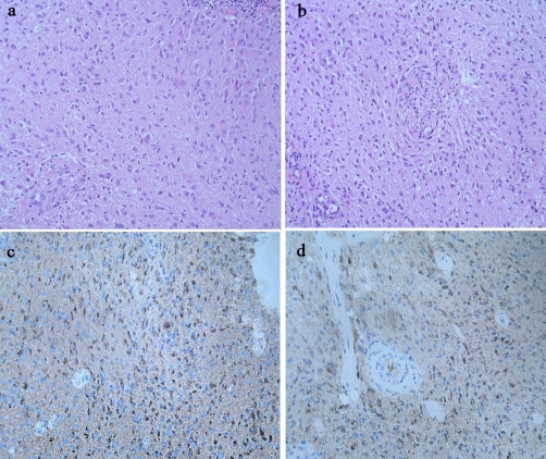
Figure 1.
Histological study of the removed masses revealed a diffuse proliferation of neoplastic spindle cells with rod-shaped nuclei infiltrating the brain parenchyma in both Patients 1 (a) and 2 (b). Paraffin-embedded sections were stained by the immunoperoxidase method and were positive for glial fibrillary acidic protein (GFAP) (c) and S100 protein (d).

