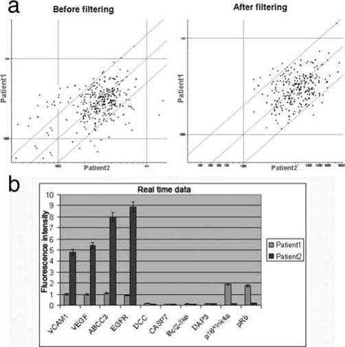
Figure 3.
(a) Scatterplot showing the gene expression difference between the two patients. Threshold lines are set to a 2-fold change. The left panel shows the data before background filtering, while the right panel shows the data after filtering. (b) Histogram showing expression data regarding the selected genes by real-time RT-PCR. Normalization was conducted on beta-actin.

