
Figure 2.
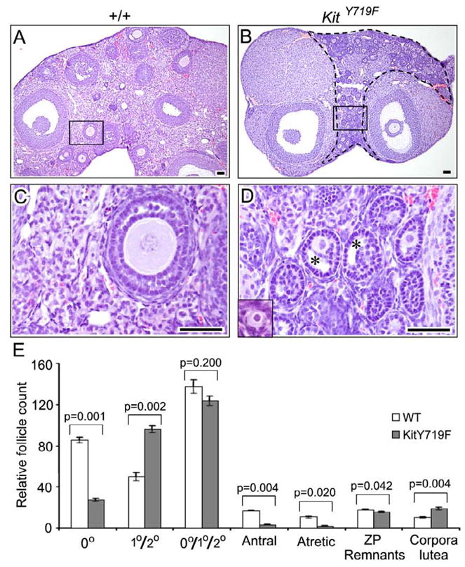
Analysis of KitY719F/KitY719F and sibling control (+/+) ovaries at 16 weeks reveals defects in primordial follicle survival/maintenance and in early follicle maturation (primary to secondary follicle transition). A–D) H&E stained tissue sections, bars = 50 microns. B) Abundance of morphologically abnormal follicles arrested at the primary/secondary follicle stage within the area enclosed by a dashed line. C–D) Higher magnification in boxed areas shown in A and B; insets show morphologically normal primordial follicles from elsewhere in each section. E) Relative follicle counts in serially sectioned ovaries (n=6 ovaries per genotype; error bars=s.e.m.). Due to the difficulty of reliably distinguishing primary from secondary follicles (because of abnormal maturation phenotype), primary + secondary follicles were counted together. P values were calculated per the student t test.