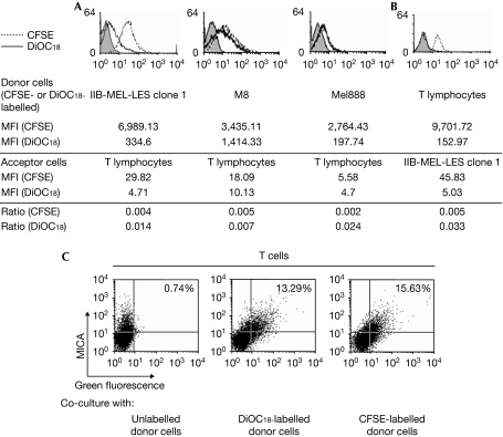
Figure 4.
Transfer of molecules between tumour and T cells. Acceptor unlabelled cells (peripheral blood mononuclear cells (PBMCs) stimulated with phytohaemagglutinin (PHA) and interleukin (IL)-2 in (A) and cells of tumour clone 1 in (B)) were co-cultured for 1 h with 3,3′-dioctadecyloxacarbocyanine perchlorate (DiOC18)- or carboxyfluorescein succinimidyl ester (CFSE)-labelled donor cells (melanoma cells in (A) or PBMCs stimulated with PHA+ IL-2 in (B)), and fluorescence was assessed on (A) non-adherent CD3+ T cells or (B) tumour cells after removing PBMCs. Numbers represent the mean fluorescent intensity (MFI) of CFSE- or DiOC18-labelled donor cells or acceptor cells that captured CFSE or DiOC18. The extent of trogocytosis was calculated as the ratio of fluorescence between acceptor and donor cells for each fluorescent probe. In addition, tumour clone 1 (unlabelled or labelled with DiOC18 or CFSE) was co-cultured for 1 h with PBMCs stimulated with PHA and IL-2, and cell surface expression of major histocompatibility complex class I chain-related protein A (MICA) was assessed by flow cytometry on non-adherent CD3+ cells (C). The percentages in quadrant 2 represent T cells that captured the fluorescent probe and MICA (quadrants were set with acceptor cells that did not contact tumour cells). Results are representative of three experiments carried out with cells from three different donors. The increase in green fluorescence on acceptor cells after co-culture with CFSE- and DiOC18-labelled donor cells (A,B) or the increase in percentage of double-positive cells after co-culture with CFSE- and DiOC18-labelled donor cells (C) was statistically significant, compared with acceptor cells cultured alone (P<0.05).

