Figure 3.
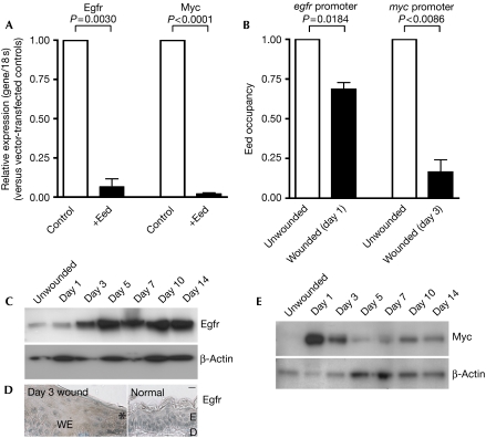
PRC2-mediated repression is decreased in the regulatory regions of important repair genes, myc and egfr, during wound healing. (A) Transfection of mIMCD3 cells with either empty vector (control) or Eed for 24 h, followed by quantitative reverse transcription–PCR analysis of Egfr and Myc, shows that they are both silenced by Eed. The results, normalized to 18 s and presented relative to levels in vector-transfected controls, show the mean±s.e.m. of at least three independent experiments (one-way ANOVA P-values indicated; n=3–5). (B) Quantitative chromatin immunoprecipitation experiments on unwounded compared with wounded skin, comparing the levels of Eed bound to egfr and myc promoter regions on days 1 and 3 post-wounding, respectively, show less Eed binding during repair. Quantitative PCR results are shown. Data are presented as mean±s.e.m. of three independent experiments (t-test P-values indicated; n=3). (C–E) Western blot analysis of Egfr and Myc during the time-course of wound repair and immunohistochemistry (D) showing increased Egfr staining (brown) in day 3 wound-edge epidermis (asterisk demarks the wound edge) compared with normal unwounded skin. Scale bar, 20 μm. ANOVA, analysis of variance; D, dermis; E, epidermis; WE, wound epithelium.

