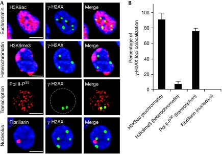
Figure 2.
γ-H2AX foci colocalize with transcribed regions in post-mitotic cells treated with camptothecin. (A) Human lymphocytes were treated with 25 μM CPT for 1 h before staining for γ-H2AX (green), and H3K9ac (euchromatin marker; red), H3K9me3 (heterochromatin marker; red), fibrillarin (nucleolus marker; red) or Pol II phosphorylated on Ser 5 (Pol II-PS5, transcription marker; red). Images were merged to determine colocalization (yellow). DNA was counterstained with DAPI (blue). Bars correspond to 5 μM. (B) Percentages of γ-H2AX foci colocalizing with H3K9ac, H3K9me3, fibrillarin or Pol II-PS5 in lymphocytes treated as in (A). Three hundred γ-H2AX foci were analysed in each group (average±standard deviation). CPT, camptothecin; DAPI, 4′,6-diamidino-2-phenylindole; γ-H2AX, phosphorylated histone H2AX; H3K9ac, acetylation of histone H3 on lysine 9; H3K9me3, trimethylation of H3K9.

