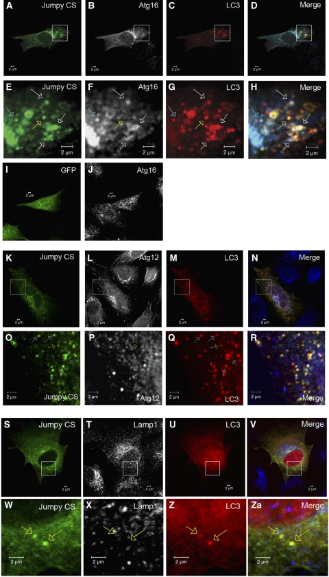
Figure 4.
Jumpy localizes to autophagic isolation membranes. (A–H) C2C12 cells were transfected for 24 h with GFP-Jumpy C330S (Jumpy CS) and tdTomato-LC3 (LC3), fixed and immunostained with anti-Atg16 (Alexa 648-labelled secondary antibody). Jumpy CS (green), LC3 (red) and Atg16 (white in B and F or blue in D and H). Boxed areas (A–D) are shown at higher magnification in the corresponding panel below (E–H). White arrows indicate colocalization among Jumpy CS, LC3 and Atg16, yellow arrow indicates colocalization between Jumpy CS and LC3 but not Atg16, blue arrow indicates colocalization between Jumpy CS and Atg16 but not LC3. (I, J) C2C12 cells were transfected for 24 h with GFP (I), fixed and immunostained with anti-Atg16 (J). (K–R) C2C12 cells were transfected for 24 h with GFP-Jumpy C330S (Jumpy CS) and tdTomato-LC3 (LC3), fixed and immunostained with anti-Atg12(M) antibody. Jumpy CS (green), LC3 (red) and Atg12 (white in L, P or blue in N, R). Boxed areas (K–N) are shown at higher magnification in the corresponding panel below (O–R). White arrows indicate colocalization among Jumpy CS, LC3 and Atg12, yellow arrow indicates colocalization between Jumpy CS and LC3 but not Atg12, blue arrow indicates colocalization between Jumpy CS and Atg12 but not LC3. (S–ZA) C2C12 cells were transfected for 24 h with Jumpy CS and Cherry-LC3 (LC3), fixed and immunostained with anti-lamp1. Jumpy CS (green), LC3 (red) and Lamp1 (white in T, X or blue in V, ZA). Boxed areas (S–V) are shown at higher magnification in the corresponding panel below (W–ZA). Yellow arrows indicate colocalization between Jumpy CS and LC3 but not Lamp1. Scale bars, 2 μm.

