Figure 2.
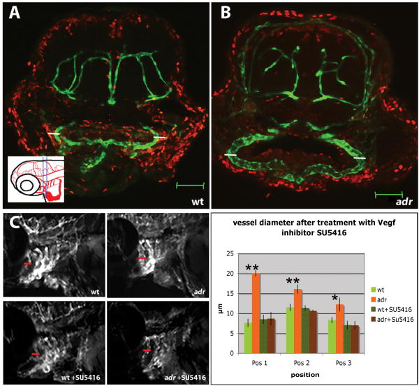
(A, B) Confocal crossections of 74hpf wild-type (A) and adrs277 mutant (B) larvae visualized for Tg(flk1:EGFP)s843 expression (green) and BrdU (red).
BrdU labeling indicates that no cell proliferation takes place between 59 and 74hpf within the endothelial cells (GFP-positive) of the 2nd visible aortic arch vessel (AA3) of wild-type (A) or adrs277 mutant (B) animals. Also note the clear dilatation of the aortic arch vessel (small white bars corresponding to 25μm in A and B). Scale bars, 50μm.
(C, D) Treatment of wild-type and adr mutant animals with the VegfR inhibitor SU5416 from 60hpf to 76hpf. (C) Epifluorescence micrographs of 76hpf untreated and SU5416 treated wild-type and adrs277 mutant larvae (red bars indicate the aortic arch diameter of untreated adr mutant larvae). (D) Diagram showing the diameter of the aortic arch vessel (AA1) in untreated and SU5416 treated wild-type and adrs277 mutant larvae. The aortic arch vessel diameter was measured at the positions indicated in Fig. 1G (4 wild-types, 3 mutants, 4-5 SU5416 treated wild-types and 3-6 SU5416 treated mutants were examined; for these latter two groups not all positions were easily visible in all embryos).

