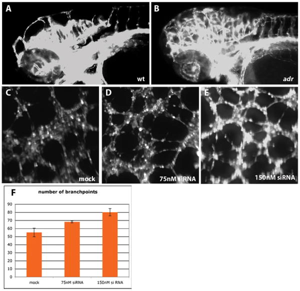
Figure 4.
Loss of Sars leads to increased sprouting in actively forming vessels.
(A, B) Aberrant vessel formation in hindbrain capillaries of adrs277 mutants (B, compare to wild-type in A). Epifluorescence micrographs of 80hpf wild-type (A) and adrs277 mutant (B) Tg(flk1:EGFP)s843 larvae, shown in lateral views. These micrographs were overexposed to reveal all the vascular branches.
(C-E) Knockdown of SARS in human endothelial cells.
Mock (C) and SARS-siRNA (D, E) transfected HUVEC's visualized by Calcein staining after a tube formation assay.
(H) Number of branchpoints in the vascular network formed by HUVECs after either mock or siRNA transfection. Shown are the mean values, error bars denote standard deviation. P<0.05 mock vs 75nM, P<0.01 mock vs 150nM, P<0.05 75nM vs 150nM.

