
Figure 6. Bdnf Overexpression Partially Rescued Dendritic Atrophy in Neurons Expressing RTT-Associated MECP2 Mutations, Increasing Dendrite Length Only to Control Levels.
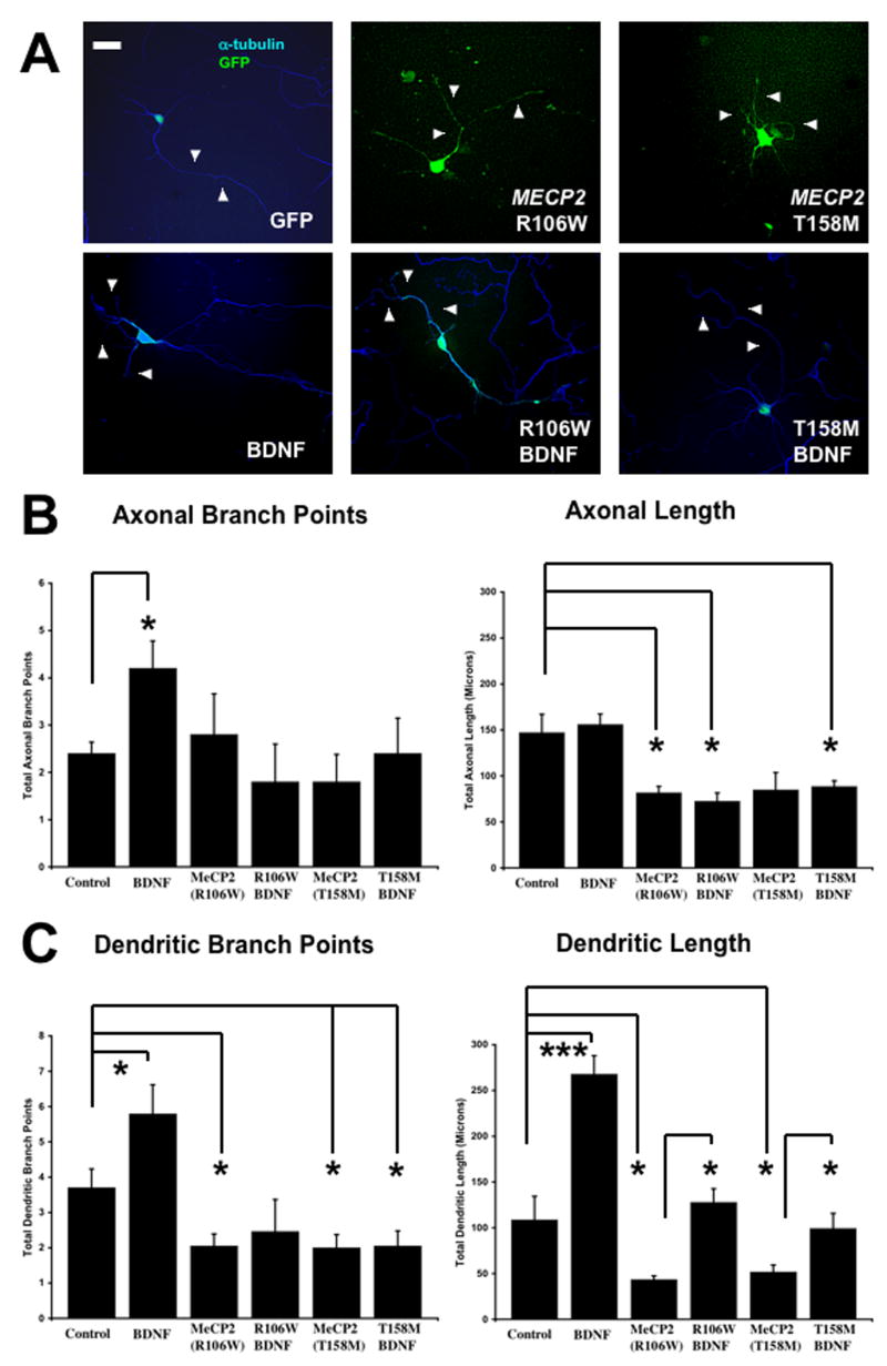
A. Representative examples of neurons transfected with a control GFP plasmid, plasmids to overexpress to different missense MECP2 mutations (R106W or T158M) commonly found in Rett syndrome patients, and a plasmid to overexpress Bdnf (scale bar = 10μm). For quantitative morphological analyses, neurons were co-transfected with eGFP (green) or stained with anti-α-tubulin antibodies (blue). B. Population data on axonal length and branch points. C. Population data on dendritic length and branch points.