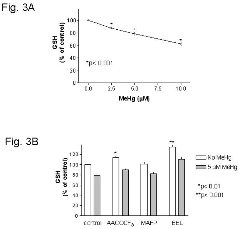
Fig. 3. Effect of PLA2inhibitors on MeHg induced GSH depletion.

Fig. 3A : Concentration dependent depletion of cellular GSH by MeHg. Mouse glia were treated with various concentrations of MeHg for 4 hours, and then the cellular GSH levels were determined. MeHg at 2.5 μM or above caused significant GSH depletion. Fig. 3B : PLA2 inhibitors did not prevent MeHg induced GSH depletion. Mouse glia were treated with 20 μM AACOCF3, 10 μM MAFP or 5 μM BEL two hours after plating. After overnight incubation, cells were treated with 5 μM MeHg (without inhibitors) for 4 hours, and then cellular GSH was determined. Two-way ANOVA analysis indicated that both MeHg and PLA2 inhibitors had significant effects on GSH levels. There was no interactions between these two treatments. Post-test Bonferroni analyses between each set of columns indicated that MeHg always caused significant (p< 0.001) GSH depletion regardless of whether cells were untreated or were treated with PLA2 inhibitors. Further analysis indicated that both AACOCF3 (p< 0.01) and BEL (p< 0.001) could significantly raise cellular GSH levels above control.
