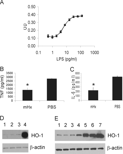
Figure 4.
Hx does not neutralize LPS as assessed by Limulus lysate actvity but acts on macrophages in a manner that does not up-regulate the expression of HO-1. (A) LAL assay of LPS preincubated with 2 μg/ml purified mHx (▴) or PBS (□). (B and C) BMDMs were preincubated with mHx (20 μg/ml) for 2 h, followed by extensive washes in serum-free medium. LPS (20 ng/ml) was then added to the culture and incubated overnight. Concentrations of TNF (B) and IL-6 (C) in the supernatants were determined by ELISA. The results represent mean ± se and are representative of five independent experiments. *, P < 0.05, compared with cells treated with PBS. (D and E) BMDMs were incubated overnight with: (D) Lane 1, PBS; Lane 2, mHx (40 μg/ml); Lane 3, rhHx (40 μg/ml); Lane 4, hemin chloride (3 μM), or (E) Lane 1, PBS; Lane 2, mHx (40 μg/ml, equal to 0.67 μM); Lane 3, mHx (200 μg/ml, equal to 3.35 μM); Lane 4, hemin-mHx complex (0.67 μM); Lane 5, hemin-mHx complex (3.35 μM); Lane 6, hemin-chloride (0.67 μM); Lane 7, hemin chloride (3 μM). Cells were then washed three times by PBS. Cell lysates were used for SDS-PAGE. Western blot was performed using anti-mouse HO-1 antibody, followed by stripping and reprobing by anti-β-actin antibody.

