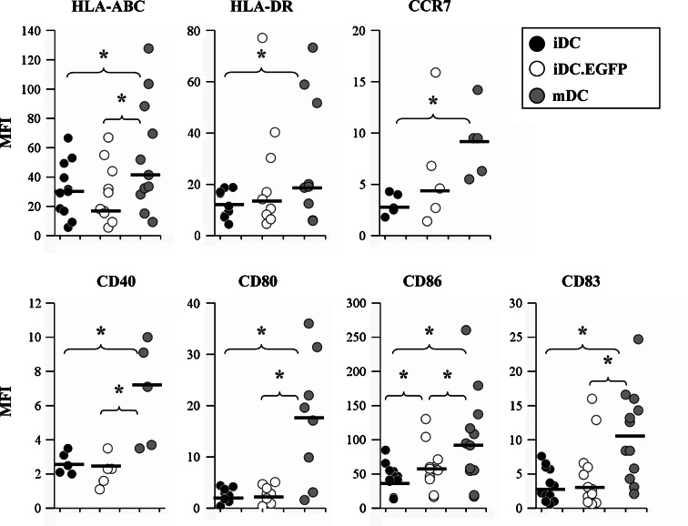
Fig. 2.
Evaluation of surface phenotypic markers expressed on DC from healthy donors. iDC, iDC.EGFP, and mDC were labeled for flow cytometry using PE-conjugated antibodies for HLA-ABC, HLA-DR, CCR7, CD40, CD80, CD86 and CD83 as described in “Materials and methods”. Data was analyzed as described in Fig. 1. Black bars represent mMFI for the donors analyzed. Asterisks represent statistically significant differences (P ≤ 0.05) between two analyzed groups according to the 2-sided paired t test

