
Figure 2.
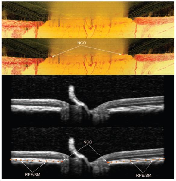
Delineation of the NCO within histomorphometric (top two images) and SD-OCT (bottom two images) reconstructions. These images are Multiview software screen captures. Top image: Digital, radial sagittal section of a histomorphometric reconstruction section (90° or horizontal in location), taken from the right eye of monkey 23540, a 9-year-old female rhesus macaque. Middle top image, red triangles: the location of the NCO (as applied within Multiview software) which in this eye is coincident with the BMO. Middle bottom image: sagittal view of a radial interpolated SD-OCT section (90° location), taken from the left eye of monkey 23511, a 12-year-old male rhesus macaque. Bottom image: the same as the middle image, but with NCO points marked (red glyphs, as applied within Multiview software). The posterior surface of the RPE/BM complex is also marked (orange lines and glyphs). The NCO points are at the innermost aspect of this surface.