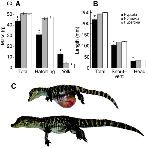
Fig. 1.
Comparison of alligator hatchlings incubated under three different oxygen levels (12%, 21% and 30%). (A) Mass measurements: total, body and yolk masses. Hypoxic hatchlings are significantly smaller than their normoxic and hyperoxic siblings, but the remaining yolk sac of hypoxic animals is significantly larger. (B) Length measurements: total, snout-to-vent and head lengths. Hypoxic hatchlings are significantly smaller than their normoxic and hyperoxic siblings. (C) A pair of anaesthetised alligator siblings, incubated under hypoxia (above) and normoxia (below). Note the diminutive hatchling size and the protruding yolk sac in the hypoxic animal. The yolk sac is completely incorporated into the abdominal cavity and the umbilical scar closed, but the abdominal skin is stretched thin and a pronounced left umbilical vein is seen. The height of the yolk sac exceeds the length of the limbs, making locomotion cumbersome. Statistical significance between groups was calculated by ANOVA with post hoc Tukey–Kramer (*P<0.05). Bar height and error bars indicate the mean ± s.e.m. for each group.

