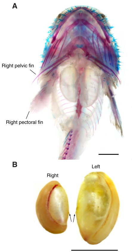
Fig. 9.
(A) Ventral view of a cleared and stained B. trispinosus specimen (6.6 cm standard length) showing the bilateral swimbladder. The skin and viscera of the abdominal cavity were removed and the swimbladder was left in place. Specimen was cleared and stained for cartilage (with Alcian Blue) and bone (with Alizarin Red) following the protocol of Song and Parenti (Song and Parenti, 1995). Scale bar represents 1 cm. The right pelvic and pectoral fins are labeled for reference orientation. (B) Ventral view of dissected swimbladders from a 9.9 cm standard length female, showing asymmetry between left and right bladders. Arrows indicate points of attachment, where the bladders are connected to each other by connective tissue. Scale bar represents 1 cm.

