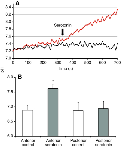
Fig. 2.
Effects of serotonin on the pHi in isolated and perfused anterior or posterior midguts. (A) pHi time courses of representative areas of interest from the anterior (red) and posterior (black) midgut of larval (4th instar) Aedes aegypti, showing the influence of addition of 0.2 μmol l–1 of serotonin (arrow) to the hemolymph-side bath. Hemolymph-side bath: oxygenated mosquito saline (pH 7). Luminal perfusate: NaCl (100 mmol l–1, pH 7). (B) Average pHi values for anterior (N=71 areas of interest from 10 different tissues) and posterior (N=19 areas of interest from four different tissues) midguts before and after addition of serotonin. Asterisk indicates statistically significant difference from control (P<0.05).

