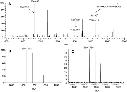
Fig. 2.
(A) Direct MALDI-FTMS spectrum of a small piece of freshly dissected Homarus americanus supraoesophageal ganglion (brain). This spectrum was measured using DHB as the matrix, with the conditions optimized for accumulation of m/z 2500. Calibration was done using the known peptide peaks, APSGFLGMRamide (CabTRP I) and VYRKPPFNGSIFamide (Val1-SIF) at m/z 934.4927 and m/z 1423.7845, respectively. As can be seen from this spectrum, a peak corresponding to that of the [M+H]+ ion for SYWKQCAFNAVSCFamide (a disulfide bridge present between the cysteine residues) was present at m/z 1650.7158 (–2.0 p.p.m. error from the theoretical m/z of 1650.7192). (B) Predicted mass and isotopic distribution for the [M+H]+ ion for SYWKQCAFNAVSCFamide (a disulfide bridge present between the cysteine residues). (C) An expansion of the measured isotopic distribution for putative SYWKQCAFNAVSCFamide, showing that the measured mass and isotopic distribution strongly support the existence of this peptide in the lobster brain.

